3D X-ray before a dental implant: when does a CBCT scan make sense?
A 3D X-ray before a dental implant is not always necessary. A standard X-ray is often enough. A cone beam CT scan (CBCT, in German DVT) helps when bone, nerves or the maxillary sinus cannot be assessed clearly enough on a two-dimensional image.
By Stela Lobzhanidze, dentist in Vienna-Meidling
Published
Key points
- Every implant starts with a clinical examination and suitable X-ray diagnostics.
- A two-dimensional X-ray is often enough, a CBCT is added when the situation is unclear or difficult.
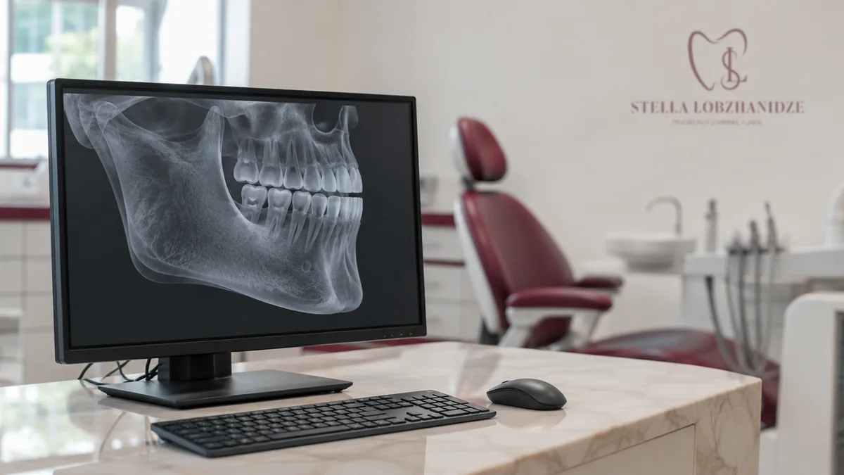
- The 3D image shows bone width, the nerve canal and the maxillary sinus without overlapping structures.
- On average, the radiation dose of a CBCT is clearly lower than that of a conventional CT, but higher than that of a small dental X-ray.
- For implant placement with a surgical guide, a CBCT is the basis of the planning.
Why do you need a 3D X-ray before a dental implant?
An implant is anchored in the jawbone. Before that, it must be clear how much bone there is and where nerves, neighbouring teeth and the maxillary sinus are located.
The German S3 guideline on 3D imaging in implant dentistry requires a clinical examination and adequate X-ray diagnostics before every implant. A two-dimensional image, such as a panoramic X-ray, is often enough. If it does not provide all the information needed, a 3D scan should be taken.
When is a standard X-ray not enough?
The guideline lists several situations in which a CBCT is useful:
- a very narrow jaw ridge or marked undercuts in the bone
- severe bone loss in the back of the lower jaw
- an unclear course of the nerve or of the roots of neighbouring teeth
- earlier diseases of or operations on the maxillary sinus
- unusual findings that need to be clarified
- planned implant placement with navigation or immediate loading
Whether you need a 3D scan is decided by your dentist after the examination. For every scan, the benefit must outweigh the radiation risk.
What does the 3D image show that a standard X-ray misses?
On a two-dimensional image, structures overlap. A CBCT shows the bone in all directions instead. This makes it easier to assess the height, width and structure of the bone.
Bone defects also become visible. This helps to decide whether bone grafting is needed before the implant.
The nerve canal in the lower jaw is usually easier to see on the 3D image than on a panoramic X-ray. Because measurements on 3D images also have small inaccuracies, a safety distance to the nerve is still planned.
How high is the radiation dose of a CBCT?
The dose depends heavily on the device and on the size of the area scanned. According to the S2k guideline on CBCT, it is on average clearly lower than that of a conventional computed tomography (CT) scan.
Compared with a small X-ray of individual teeth, however, the dose is at least ten times higher. That is why only the area actually needed for the question is scanned.
For children and teenagers the decision is weighed especially carefully. Pregnancy is a reason to be very cautious. Please tell us beforehand if you are or might be pregnant.
What happens during a CBCT scan?
During the scan, the device rotates once around your head. On average this takes about 8 to 20 seconds.
Keep as still as possible during this time. Movement blurs the image and can mean the scan has to be repeated.
Metal in the mouth, such as metal crowns or fillings, can cause streaks on the image. Your dentist takes this into account when reading the scan.
How is the 3D image used for planning with a surgical guide?
In computer-assisted implant placement, the position of the implant is planned on screen. From this, a surgical guide is made that sets the drilling direction during the procedure. The basis is a CBCT, combined with an impression or scan of the teeth.
According to the current S3 guideline, guided implants deviate less from the plan on average than implants placed freehand. Small deviations are still possible. Whether a guide makes sense is decided together with you.
Where can you get a CBCT before an implant in Vienna?
Our practice in 1120 Vienna-Meidling has its own CBCT device. We use 3D scans mainly for planning implants and surgical procedures.
Before any treatment you receive a detailed cost estimate. Book an appointment on +43 1 813 31 23 or online at stelladent.termin365.at.
Book an appointment
Book online or give us a call.
Sources
- DGZMK/DGI – S3-Leitlinie Implantologische Indikationen für die Anwendung von 3D-Röntgendiagnostik und Navigation (2021)
- DGZMK – S2k-Leitlinie Dentale digitale Volumentomographie (2022)
- DGI/DGZMK – S3-Leitlinie Konventionelle und computerassistierte Implantatinsertion (2025)
- gesundheit.gv.at – Zahnimplantate
- gesundheit.gv.at – Strahlenanwendung in der Medizin
This article is for general information only and does not replace a dental examination or advice.